  • Image
    Patienten

Closed Loop Stimulation

Myocardial Contractility Measurement

CLS, the market's only myocardial contractility measurement, has benefitted patients for more than 25 years. CLS translates beat-to-beat myocardial contractility variations into physiologic pacing rate changes that appropriately support metabolic demand.

By integrating with the body's autonomic nervous system, CLS puts the body back in control of heart rate, rather than secondary or tertiary measures like breathing or motion.

CLS is available in all BIOTRONIK pacemakers, ICDs and CRT devices. 

Product Highlights

Image
Icon

11% increase in patient performance during daily activities compared to accelerometers.¹

Image
Icon

16% lower risk for first AHRE > 6 minutes, Stroke or TIA vs. DDDR accelerometer²

Image
Icon

69% decrease in risk of AT/AF occurrence.³

Overview

Why would your patients need a comprehensive physiologic pacing?

Every person experiences physical and emotional stress during their life. Regular daily activities, sport, or even playing chess or watching a video can result in such stress. If they suffer from chronotropic incompetence (CI), the patient's heart is not able to respond correctly to the stimulus. Studies have shown that CLS can improve the quality of life of CI patients as well as those suffering from vasovagal syncope.4

Thanks to CLS rate adaptation, pacemaker patients are also supported during emotional stress.

Which patients can benefit from CLS?

CLS is beneficial for patients with Chronotropic Incompetence (CI) and Vasovagal Syncope (VS) as well as patients with Atrial Fibrillation (AF). For patients suffering from CI, the PROVIDE study showed that 75% of patients programmed with DDD-CLS experienced significant improvement compared to only 22% of patients programmed in DDDR.5

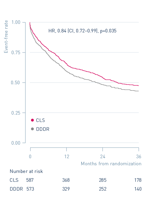
For patients burdened with paroxysmal or permanent atrial fibrillation (AF), cardiac output is also of utmost importance when AV synchrony is lost. CLS is able to support these patients through a steady, reliable and flexible algorithm. CLS has also been shown to reduce the burden for atrial tachycardia patients compared with other modes. In the BURDEN III trial, CLS showed a 16% risk reduction of the primary endpoint of time to first atrial high-rate episode of at least six minutes, stroke or TIA compared to accelerometer. That risk reduction to 23% for patients with no AV block and 27% for patients with no history of AF. 

For patients suffering from VS, the recurrence of syncope significantly decreases with a CLS system. The randomized, placebo-controlled, double-blinded, multicenter BIOSync CLS study demonstrated a reduction of syncope recurrence rate after two years by 77% with CLS pacing versus placebo.6

CLS continuously translates variations in myocardial contractility into physiologic pacing rate changes to appropriately support metabolic demands

Primary Sensor (insert reference): Take measurements directly from myocardial tissue

Built On A Beat-To-Beat Architecture (insert reference): Rapidly evaluates every beat, and is able to respond on the next beat

Automated With Machine Learning (insert reference): Learns the patient while automatically recalibrating to systemic variations (changes in medication, hydration, etc.)

Additional technical details

Step 1: Creation of the rest curve

  • CLS measures the intracardiac impedance throughout each ventricular contraction. 
  • When the patient is at rest, reference curves are created by using the average of the last 256 curves of the paced and sensed events. This reference is continuously adapted to the actual resting conditions. 

How is the impedance curve created?

The impedance is measured at the tip of the right ventricular lead and it reflects changes seen during systole.

At the beginning of systole, blood and muscle volume define the impedance value (1). When the contraction starts, the percentage of muscle volume will increase, resulting in a higher impedance value (2) (3) (4).

Step 2: Comparing the rest curve with the load curve

  • For each heartbeat, CLS determines the impedance curve during the ventricular contraction and compares it to its resting reference curve. No special leads or fixation positions are needed for measuring contraction. 
  •  Slight change between curves will result in a minimum change in rate adaption.The same applies to medium and large changes.

Step 3: Automatic adjustements of CLS

  • CLS is initialized during the auto-initialization phase of the cardiac device. 
  • CLS is automated with machine learning and responds to the patient on a beat to beat basis.
  • Changes in patients’ drug therapy or cardiac remodeling does not affect the rate adaption as it automatically updates the curves with the new data. 

Media

Testimonials

Unique to BIOTRONIK pacemakers, Closed Loop Stimulation (CLS) sensor technology offers the most physiological rate regulation on the market.
Dr Prasad shares his experiences and opinion regarding the BIOTRONIK's CLS sensor compared with the accelerometer.
Based on his experience, Dr Dayer gives his opinion regarding the BIOTRONIK's CLS sensor compared with the accelerometer.
Janine, a 36-year-old teacher, was diagnosed with vasovagal syncope. Thanks to a BIOTRONIK device with CLS, she was able to return to a normal life and resume doing what she loves most: teaching.

Contact Us

I prefer to be contacted by
*Fields are mandatory.

References

1. Abi-Samra FM, Singh N, Rosin BL, Dwyer JV, and Miller C. Europace. 2013; 15: 849-856. 2. Pisano E, et al. Closed loop stimulation reduces the incidence of atrial high-rate episodes compared with conventional rate-adaptive pacing in patients with sinus node dysfunctions, EP Europace, Volume 26, Issue 7, July 2024, euae175. 3. Ikeda S, Nogami A, Inoue K, et al. Closed‐loop stimulation as a physiological rate‐modulated pacing approach based on intracardiac impedance to lower the atrial tachyarrhythmia burden in patients with sinus node dysfunction and atrial fibrillation. J Cardiovasc Electrophysiol. 2020; 31: 1187-1194. 4. E. Occhetta et al., Closed loop stimulation in prevention of vasovagal syncope. Inotropy controlled pacing in vasovagal syncope (INVASY). 5. Pavri BB et al., An Impedance Sensor (CLS) is Superior to Accelerometer for Chronotropically Incompetent Patients with Sinus Node Dysfunction: Results of a Pilot Study with a Dual Sensor Pacemaker 6. Brignole M. et al., Cardiac pacing in severe recurrent reflex syncope and tilt-induced asystole