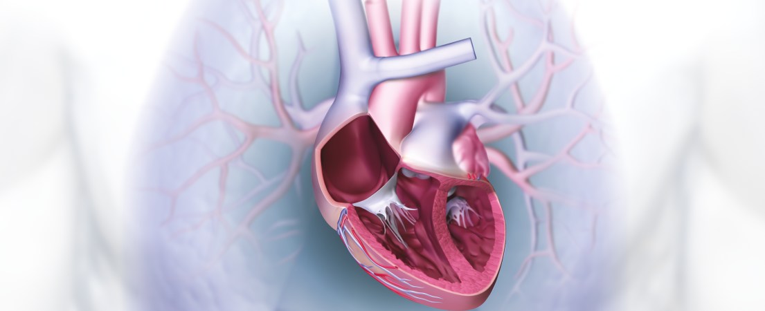
The human Heart
The Heart – The Engine of Life
The circulatory system carries vital oxygen, water, nutrients and hormones through our bodies. Our “engine” – the heart – keeps this system running. The fist-sized muscle weighs 300 grams (10.5 ounces) and is located in the center of the chest, slightly to the left, protected by the breastbone (sternum) and ribs. In the span of one minute, the heart pumps all of our blood through the entire body with rhythmic contractions. Over one’s lifetime, the heart beats three billion times and transports 66 million gallons of blood – an astonishing performance that no other engine can compete with.
Two Separate Circulatory Systems
The heart has two halves that beat in the same rhythm. Each half is divided into an upper chamber (the atria) and a lower chamber (the ventricles). These sides supply two different circulatory systems. The right side carries blood that is low in oxygen from the body to the right atrium and into the lungs. There, the blood takes in oxygen and releases carbon dioxide. The left side of the heart transports oxygenated blood from the lungs into the left atrium and the rest of the body, including the head, arms and legs and other organs. Valves that open and close the heart’s chambers regulate blood flow.
The heart’s halves differ in size because they support separate circulatory systems. The left half is bigger and surrounded by a strong layer of muscle because it works harder pumping oxygenated blood throughout the body.
The Sinus Node Sets the Heartbeat
Two sounds that can be heard with a stethoscope make up the heartbeat. One sound – the systole phase – is the heart chambers contracting and pumping blood into both circulatory systems. This lasts about one third of a second. The diastole phase – the second sound – is the atria contracting when the lower heart chambers are empty. It lasts approximately two thirds of a second. When the atria are full, the lower chambers are empty, and vice versa.
The sinus node, a plexus in the right atrium, starts the contractions. From there, the impulse travels into the chambers. These electrical currents can be seen with an electrocardiogram (ECG or EKG). The physician uses the ECG to evaluate if the heart is healthy and working well, and see if the patient has previously suffered a heart attack.
The brain and nervous system control the heart. The electrical impulses that create the heartbeat come from the heart itself. The heart will beat as long as it is supplied with oxygen, even if that oxygen is supplied externally, for example during a heart transplant.
Pacemakers Fix an Irregular Heart Rhythm
Occasional extra heartbeats, called extrasystoles, do not usually need treatment. If the heart rhythm is consistently irregular, for example if it is too slow or pauses (link to bradycardia), a pacemaker can help. Implanted pacemakers were invented in the late 1950s and are continuously being improved. Today’s devices are just a little bigger than a silver dollar and their batteries last for years. They are inserted below the collarbone or above the chest muscle and connected to the heart with electrodes. Pacemakers monitor the heart’s activity and give an electrical impulse if the heart skips a beat or slows down.
Modern pacemakers like BIOTRONIK devices with Closed Loop Stimulation are also able to regulate the heart rate during physical or acute mental stress. ProMRI pacemakers allow patients to safely undergo MRI scans.

